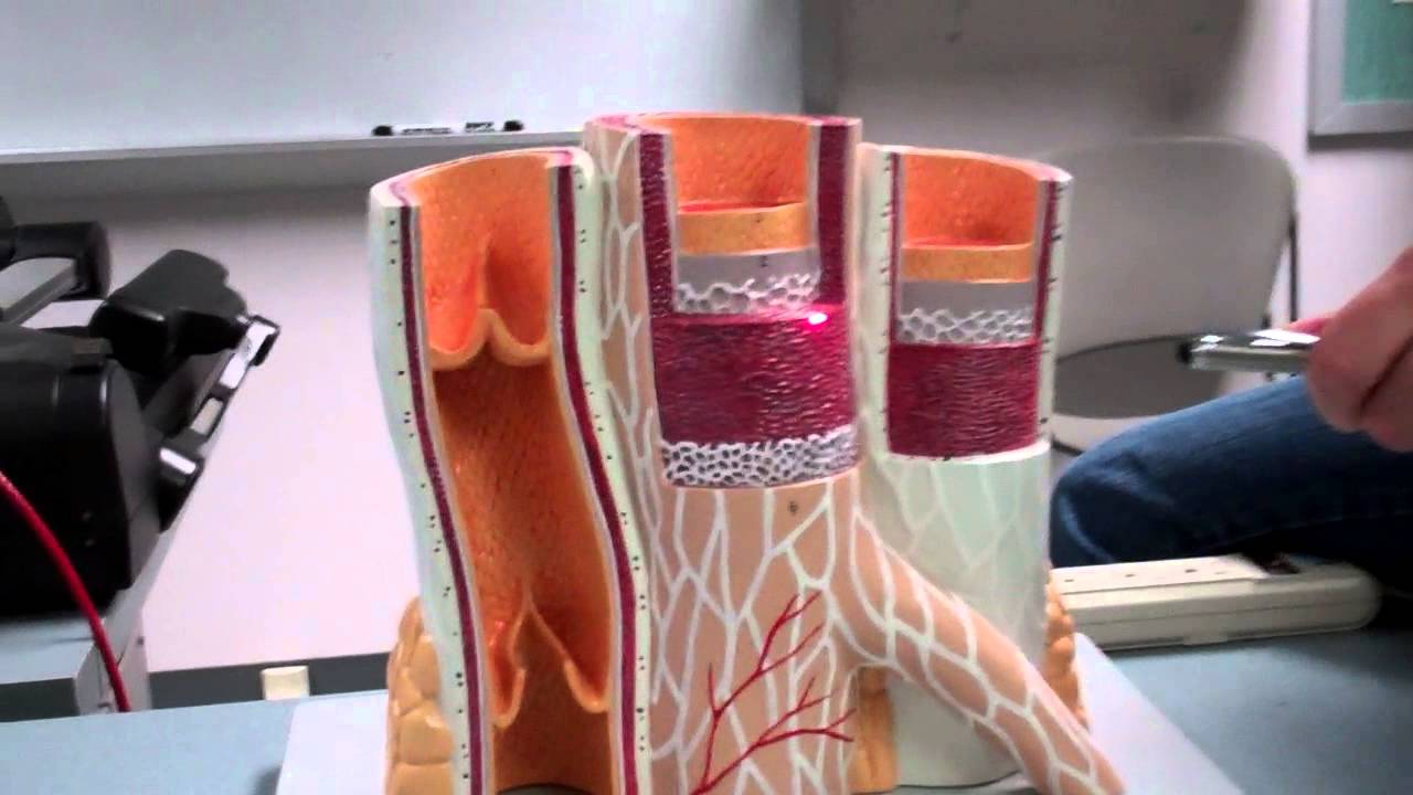
Blood Vessels Labeled / Anatomy of the Abdominal Blood Vessels | Doctor Stock / Dimitrios mytilinaios md, phd • last reviewed:. 03.12.2019 · labeled diagram showing the structure of a blood vessel. The central opening of a blood vessel, the lumen, is surrounded by a wall consisting of three layers: They include arteries, veins, and capillaries. Arteries carry blood away from the heart. Blood vessels cannot function properly when inhibited by vascular diseases.
This is an online quiz called blood vessel labeling. To view these resources with no ads, please login or subscribe (and help support our site). Blood vessels consist of arteries, arterioles, capillaries, venules, and veins. When chemoreceptors in blood vessels detect high levels of carbon dioxide in the blood, they stimulate all of the following changes except. They are vital for carrying nutrients, oxygen and waste around the body.
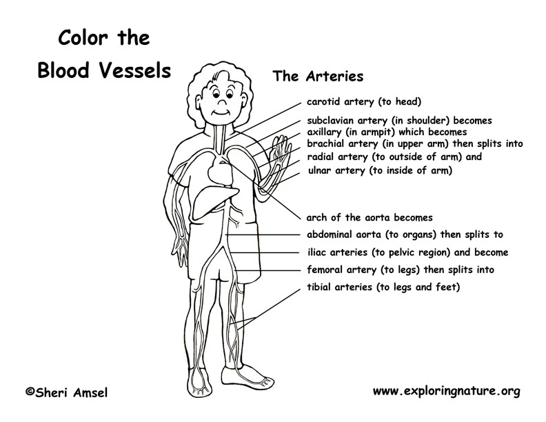
Vascular System Models - Arteries, Veins, Blood Cells ... from s-media-cache-ak0.pinimg.com Blood vessels are intricate networks of hollow tubes that transport blood throughout the entire body. Blood vessels (labeled) coloring page. Blood vessels flow blood throughout the body. Blood vessels are flexible tubes that carry blood, associated oxygen, nutrients, water, and hormones throughout the body. Allows diffusion of gases and nutrients from blood into the body cells. Carry blood towards the heart (usually deoxygenated blood, except for the pulmonary vein). Does not cover the pathology content. National institute of diabetes and digestive and kidney diseases, national institutes of health.
The capillaries also connect the branches of arteries and to.
Blood, the heart and the vessels that carry blood around the body together make up the cardiovascular system. They also take waste and carbon dioxide away from the tissues. Does not form part of the actual practical class based upon the virtual slides. Blood vessels are intricate networks of hollow tubes that transport blood throughout the entire body. Blood vessels are flexible tubes that carry blood, associated oxygen, nutrients, water, and hormones throughout the body. Pictures and 3d models played a great role in helping me learn anatomy. National institute of diabetes and digestive and kidney diseases, national institutes of health. If a blood vessel breaks, tears, or is cut, blood leaks out, causing bleeding. Blood vessels consist of arteries, arterioles, capillaries, venules, and veins. • identification of blood vessels as arteries, capillaries or veins from the structure of their walls. In successful embryos, dii labeled blood vessels are present throughout the. This is an online quiz called blood vessel labeling. One of the most common diseases of the arteries is called atherosclerosis.
All blood vessels are specifically structured to perform their function. Deep veins, located in the center of the leg near the leg bones, are enclosed by muscle. The blood vessels are the components of the circulatory system that transport blood throughout the human body. Molly smith dipcnm, mbant • reviewer: Hma practical 3 for monday july 23 and wednesday july 25.
Blood Vessels (Labeled) Coloring Page from www.exploringnature.org The tunica intima is the inner layer facing the blood. Veins (in blue) are the blood vessels that return blood to the heart. 10 photos of the the human blood vessels labeled. Hma practical 3 virtual slides. The difference in the structural characteristics of arteries, capillaries and veins is attributable to their respective functions. Blood flows throughout the body tissues in blood vessels, via bulk flow (i.e., all constituents together and in one direction). 2,731 blood vessels labeling machine products are offered for sale by suppliers on alibaba.com, of which labeling machines accounts for 5%. Nutrients and metabolic end products move between the capillary vessels and the surroundings of the cell through the interstitial fluid by diffusion and mediated transport.
As a medical student, i found anatomy pretty challenging.
Deep veins, located in the center of the leg near the leg bones, are enclosed by muscle. Three types of blood vessels that make up the entire system. Allows diffusion of gases and nutrients from blood into the body cells. These large vessels have tiny arteries, capillaries and veins in their tunica externa. This is an online quiz called blood vessel labeling. When chemoreceptors in blood vessels detect high levels of carbon dioxide in the blood, they stimulate all of the following changes except. To view these resources with no ads, please login or subscribe (and help support our site). Hma practical 3 for monday july 23 and wednesday july 25. • identification of blood vessels as arteries, capillaries or veins from the structure of their walls. They also take waste and carbon dioxide away from the tissues. Vascular injection only labels luminised vessels and therefore does not identify unopened capillaries, endothelial tip cells or isolated endothelial. One of the most common diseases of the arteries is called atherosclerosis. It is composed of an innermost layer of endothelium (simple squamous epithelium) surrounded by variable amounts of connective tissues.
Blood vessels consist of arteries, arterioles, capillaries, venules, and veins. 03.12.2019 · labeled diagram showing the structure of a blood vessel. 2,731 blood vessels labeling machine products are offered for sale by suppliers on alibaba.com, of which labeling machines accounts for 5%. They also take waste and carbon dioxide away from the tissues. Model labeled labeled body arteries cat dissection blood vessels blank blood vessel diagram exercise 32 anatomy of blood vessels capillary blood vessel blood vessel drawing label the major blood vessels blood vessel structure cardiovascular system blood vessels blood.
Blood Vessel Histology Model - YouTube from i.ytimg.com Carry blood towards the heart (usually deoxygenated blood, except for the pulmonary vein). Hma practical 3 for monday july 23 and wednesday july 25. Vasa vasorum is the blood supply of the larger blood vessels themselves, such as the aorta. Blood flows throughout the body tissues in blood vessels, via bulk flow (i.e., all constituents together and in one direction). Blood vessels labeled simple : Blood vessels 2 labeled palmar arch digital artery right femoral a right femoral v great saphenous vein left popliteal a right anterior tibial a. Blood vessels (labeled) coloring page. Master blood vessels with diagrams and arteries and veins quizzes:
Observe the blood vessels diagrams above, where you can see the structures of arteries and veins clearly labeled.
03.12.2019 · labeled diagram showing the structure of a blood vessel. The central opening of a blood vessel, the lumen, is surrounded by a wall consisting of three layers: It is composed of an innermost layer of endothelium (simple squamous epithelium) surrounded by variable amounts of connective tissues. Does not cover the pathology content. Blood flows throughout the body tissues in blood vessels, via bulk flow (i.e., all constituents together and in one direction). Embryo (figure 3c,d) including capillary beds (figure 3d). One of the most common diseases of the arteries is called atherosclerosis. Model labeled labeled body arteries cat dissection blood vessels blank blood vessel diagram exercise 32 anatomy of blood vessels capillary blood vessel blood vessel drawing label the major blood vessels blood vessel structure cardiovascular system blood vessels blood. Dimitrios mytilinaios md, phd • last reviewed: I tried to come up w a new way of teaching. Torso with the heart and blood vessels labeled. Does not form part of the actual practical class based upon the virtual slides. To view these resources with no ads, please login or subscribe (and help support our site).